Home

Refrain bâtiment leur pdb base Chine À laube Pâturer

RCSB PDB - 1QC0: CRYSTAL STRUCTURE OF A 19 BASE PAIR COPY CONTROL RELATED  RNA DUPLEX
RCSB PDB - 1QC0: CRYSTAL STRUCTURE OF A 19 BASE PAIR COPY CONTROL RELATED RNA DUPLEX

RCSB PDB - 5HNQ: Base Pairing and Structure Insights into the  5-Formylcytosine in RNA Duplex
RCSB PDB - 5HNQ: Base Pairing and Structure Insights into the 5-Formylcytosine in RNA Duplex

RCSB PDB - 3EDE: Structural base for cyclodextrin hydrolysis

RCSB PDB - 145D: Structure and thermodynamics of nonalternating C/G base  pairs in Z-DNA: the 1.3 angstroms crystal structure of the asymmetric  hexanucleotide D(M(5)CGGGM(5) CG)/D(M(5)CGCCM(5)CG)
RCSB PDB - 145D: Structure and thermodynamics of nonalternating C/G base pairs in Z-DNA: the 1.3 angstroms crystal structure of the asymmetric hexanucleotide D(M(5)CGGGM(5) CG)/D(M(5)CGCCM(5)CG)

RCSB PDB - 3O82: Structure of BasE N-terminal domain from Acinetobacter  baumannii bound to 5'-O-[N-(2,3-dihydroxybenzoyl)sulfamoyl] adenosine
RCSB PDB - 3O82: Structure of BasE N-terminal domain from Acinetobacter baumannii bound to 5'-O-[N-(2,3-dihydroxybenzoyl)sulfamoyl] adenosine
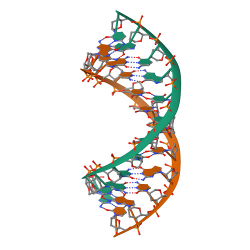
RCSB PDB - 1P43: REVERSE PROTONATION IS THE KEY TO GENERAL ACID-BASE  CATALYSIS IN ENOLASE
RCSB PDB - 1P43: REVERSE PROTONATION IS THE KEY TO GENERAL ACID-BASE CATALYSIS IN ENOLASE

RCSB PDB - 1EC4: SOLUTION STRUCTURE OF A HEXITOL NUCLEIC ACID DUPLEX WITH  FOUR CONSECUTIVE T:T BASE PAIRS
RCSB PDB - 1EC4: SOLUTION STRUCTURE OF A HEXITOL NUCLEIC ACID DUPLEX WITH FOUR CONSECUTIVE T:T BASE PAIRS

RCSB PDB - 1GQU: Crystal structure of an alternating A-T oligonucleotide  fragment with Hoogsteen base pairing
RCSB PDB - 1GQU: Crystal structure of an alternating A-T oligonucleotide fragment with Hoogsteen base pairing
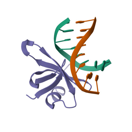
RCSB PDB - 8DH4: T7 RNA polymerase elongation complex with unnatural base  dPa-DsTP pair
RCSB PDB - 8DH4: T7 RNA polymerase elongation complex with unnatural base dPa-DsTP pair

RCSB PDB - 225D: A TETRAMERIC DNA STRUCTURE WITH PROTONATED  CYTOSINE:CYTOSINE BASE PAIRS
RCSB PDB - 225D: A TETRAMERIC DNA STRUCTURE WITH PROTONATED CYTOSINE:CYTOSINE BASE PAIRS

RCSB PDB - 7SDF: [C:Ag+:S] Metal-mediated DNA base pair in a  self-assembling rhombohedral lattice
RCSB PDB - 7SDF: [C:Ag+:S] Metal-mediated DNA base pair in a self-assembling rhombohedral lattice

RCSB PDB - 2FZA: Crystal structure of d(GCGGGAGC): the base-intercalated  duplex
RCSB PDB - 2FZA: Crystal structure of d(GCGGGAGC): the base-intercalated duplex

RCSB PDB - 1C8C: CRYSTAL STRUCTURES OF THE CHROMOSOMAL PROTEINS SSO7D/SAC7D  BOUND TO DNA CONTAINING T-G MISMATCHED BASE PAIRS
RCSB PDB - 1C8C: CRYSTAL STRUCTURES OF THE CHROMOSOMAL PROTEINS SSO7D/SAC7D BOUND TO DNA CONTAINING T-G MISMATCHED BASE PAIRS
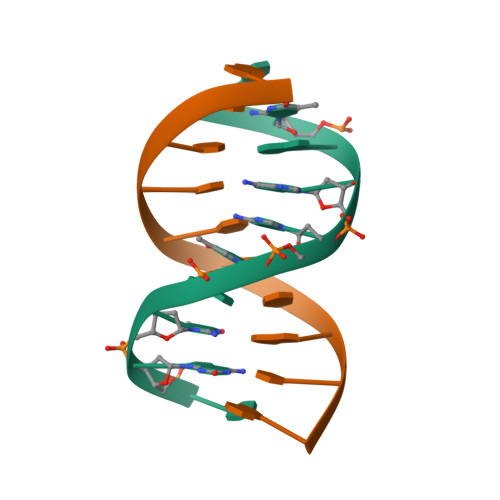
RCSB PDB - 1BE5: STRUCTURAL STUDIES OF A STABLE PARALLEL-STRANDED DNA  DUPLEX INCORPORATING ISOGUANINE:CYTOSINE AND ISOCYTOSINE:GUANINE BASE PAIRS  BY NMR, MINIMIZED AVERAGE STRUCTURE
RCSB PDB - 1BE5: STRUCTURAL STUDIES OF A STABLE PARALLEL-STRANDED DNA DUPLEX INCORPORATING ISOGUANINE:CYTOSINE AND ISOCYTOSINE:GUANINE BASE PAIRS BY NMR, MINIMIZED AVERAGE STRUCTURE
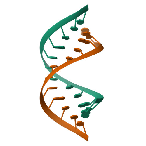
RCSB PDB - 255D: CRYSTAL STRUCTURE OF AN RNA DOUBLE HELIX INCORPORATING A  TRACK OF NON-WATSON-CRICK BASE PAIRS
RCSB PDB - 255D: CRYSTAL STRUCTURE OF AN RNA DOUBLE HELIX INCORPORATING A TRACK OF NON-WATSON-CRICK BASE PAIRS

RCSB PDB - 3EDD: Structural base for cyclodextrin hydrolysis

RCSB PDB - 1KBD: SOLUTION STRUCTURE OF A 16 BASE-PAIR DNA RELATED TO THE  HIV-1 KAPPA B SITE
RCSB PDB - 1KBD: SOLUTION STRUCTURE OF A 16 BASE-PAIR DNA RELATED TO THE HIV-1 KAPPA B SITE

RCSB PDB - 2N5O: Universal Base oligonucleotide structure

RCSB PDB - 1LCD: STRUCTURE OF THE COMPLEX OF LAC REPRESSOR HEADPIECE AND AN  11 BASE-PAIR HALF-OPERATOR DETERMINED BY NUCLEAR MAGNETIC RESONANCE  SPECTROSCOPY AND RESTRAINED MOLECULAR DYNAMICS
RCSB PDB - 1LCD: STRUCTURE OF THE COMPLEX OF LAC REPRESSOR HEADPIECE AND AN 11 BASE-PAIR HALF-OPERATOR DETERMINED BY NUCLEAR MAGNETIC RESONANCE SPECTROSCOPY AND RESTRAINED MOLECULAR DYNAMICS
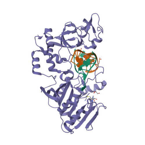
RCSB PDB - 2IH2: Crystal structure of the adenine-specific DNA  methyltransferase M.TaqI complexed with the cofactor analog AETA and a 10  bp DNA containing 5-methylpyrimidin-2(1H)-one at the target base partner  position
RCSB PDB - 2IH2: Crystal structure of the adenine-specific DNA methyltransferase M.TaqI complexed with the cofactor analog AETA and a 10 bp DNA containing 5-methylpyrimidin-2(1H)-one at the target base partner position
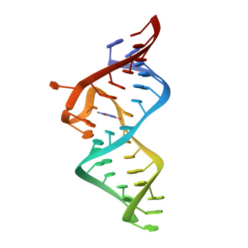
RCSB PDB - 1O15: THEOPHYLLINE-BINDING RNA IN COMPLEX WITH THEOPHYLLINE,  NMR, REGULARIZED MEAN STRUCTURE, REFINEMENT WITH TORSION ANGLE AND BASE-BASE  POSITIONAL DATABASE POTENTIALS AND DIPOLAR COUPLINGS
RCSB PDB - 1O15: THEOPHYLLINE-BINDING RNA IN COMPLEX WITH THEOPHYLLINE, NMR, REGULARIZED MEAN STRUCTURE, REFINEMENT WITH TORSION ANGLE AND BASE-BASE POSITIONAL DATABASE POTENTIALS AND DIPOLAR COUPLINGS

RCSB PDB - 2BR0: DNA Adduct Bypass Polymerization by Sulfolobus  solfataricus Dpo4. Analysis and Crystal Structures of Multiple Base-Pair  Substitution and Frameshift Products with the Adduct 1,N2-Ethenoguanine
RCSB PDB - 2BR0: DNA Adduct Bypass Polymerization by Sulfolobus solfataricus Dpo4. Analysis and Crystal Structures of Multiple Base-Pair Substitution and Frameshift Products with the Adduct 1,N2-Ethenoguanine
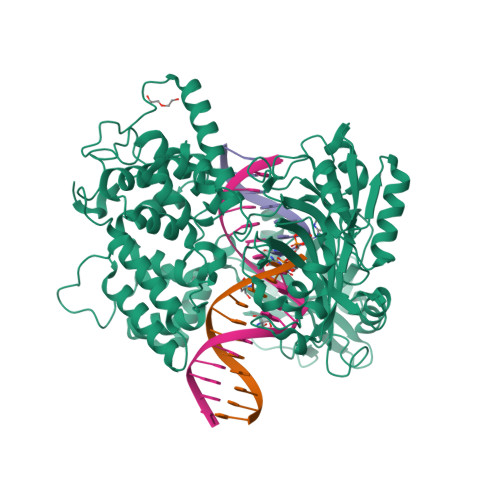
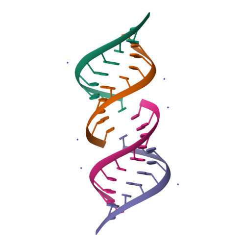
RCSB PDB - 6P0E: Human DNA Ligase 1 (E346A,E592A) bound to adenylated DNA  containing an 8-oxo guanine:adenine base-pair
RCSB PDB - 6P0E: Human DNA Ligase 1 (E346A,E592A) bound to adenylated DNA containing an 8-oxo guanine:adenine base-pair